Farm introduction
The case presented on a multi site farm of 2000 females located in Chile, with weekly production flows. The breeders are located within 30 kilometers of Site 2 and 3 (see Figure 1), the latter are separated and in order to enter, each take appropriate biosecurity measures. The finishing phase begins at approximately 75 days of age and pigs are placed in a feedlot site consisting of 15 building with a deep bedding system using wheat straw, each building has the capacity for 1150 fattening pigs which are sold to 170 days of age. The feed is manufactured by the company. The farm has a controlled health status and their vaccination plan is implemented upon weaning, using the Circovirus and Mycoplasma vaccines. The finishing phase production unit is located in a geographical area of low pig density

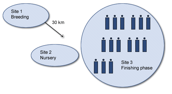
Figure 1. Geographical distribution of farm facilities.
Emergence of the case
In July 2011, the owner of the company contacted the country’s diagnostic pig laboratory, because the slaughterhouse’s Veterinarian Inspector informed that about 75% of slaughtered pigs had their livers discarded due to lesions characteristic of parasitic migration. The following week, a diagnostic laboratory employee was responsible for personally visiting the slaughterhouse and conducted an inspection of the livers of 190 animals and about a week later the same procedure was performed with the same number of pigs. In addition, he also asked the veterinary inspector how long he had been seeing this problem, to which he responds that the farm had been characterized as positive for scarring lesions due to parasite migration for a long time, with rates not greater than 5%. However, he said that the number of discarded livers had increased significantly in some batches of pigs over the last few weeks. The results of the inspection can be seen in Table 1.
Table 1. Results of pigs analyzed in the slaughterhouse.
| Week 1 | |
| Pigs tested | 190 |
| Discarded livers | 140 |
| Prevalence of scarred livers | 74% |
| Week 2 | |
| Pigs tested | 192 |
| Discarded livers | 54 |
| Prevalencia hígados cicatrizados | 28% |
After the evaluation it was determined that many pigs had lesions characteristic of parasite migration in the liver. Another thing that caught my attention was that the prevalence from one week to another was significantly different. The livers had mild scarring due to parasite migration, although none exceeded 4 scars (Figure 2 and 3). In some cases scars could be seen in the gallbladder (Figure 4).

Figure 2. Scar lesion on liver due to parasite migration
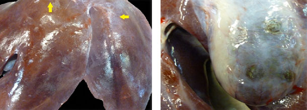
| Figure 3. Scarring lesions on liver due to parasite migration. |
Figure 4. Scarring lesions on gallbladder due to parasite migration. |
Determination of infestation dynamics within the productive system
In order to determine the dynamics of the Ascaris suum infestation, samples were taken to determine at what age animals were becoming infestated and to come up with a treatment strategy.
Ascaris suum infestations are mainly diagnosed by fecal analysis. Feces are taken directly from the rectum, trying to keep the fecal matter moist (Figure 5).

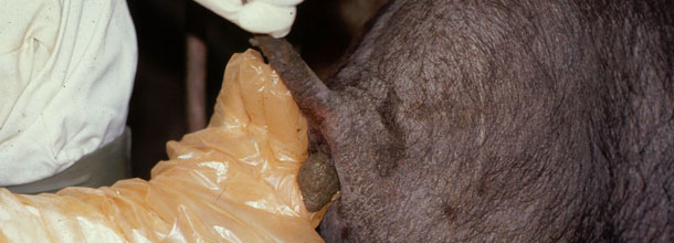
Figure 5. Fecal sampling, the contents of feces remain in the same glove and are sent to the laboratory refrigerated. The glove should not have talc.
Sampling scheme
For sampling at the farm, the following scheme was established:
- 5 samples from each finishing building.
Each sample was taken at random and recorded the average age of the pigs per building.
Coproparasitary examination Results
Once samples are taken, these were referred to the Swine Diagnostic Laboratory, where a coproparasitary examination was performed using the flotation technique with subsequent use of lens. The results were as follows:
| Building | Number of samples | Average age / building | Result |
| Building 1 | 5 | 152 days | Negative |
| Building 2 | 5 | 159 days | Negative |
| Building 3 | 5 | 166 days | Negative |
| Building 4 | 5 | 169 days | Negative |
| Building 5 | 5 | 75 days | Negative |
| Building 6 | 5 | 83 days | Negative |
| Building 7 | 5 | 92 days | Negative |
| Building 8 | 5 | 101 days | Negative |
| Building 9 | 5 | 109 days | Negative |
| Building 10 | 5 | 117 days | Negative |
| Building 11 | 5 | 123 days | Negative |
| Building 12 | 5 | 129 days | Negative |
| Building 13 | 5 | 137 days | Negative |
| Building 14 | 5 | 145 days | Negative |
Interpretation of results
Surprisingly, the results of samples tested using the coproparasitary flotation technique were negative for an eggs count. No pig was positive.
Farm Action Plan
After the analysis of laboratory results they consulted on how to best control Ascaris suum, the answer was:
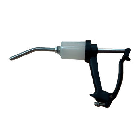
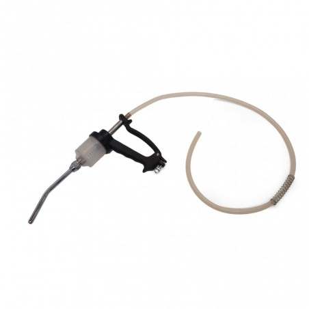
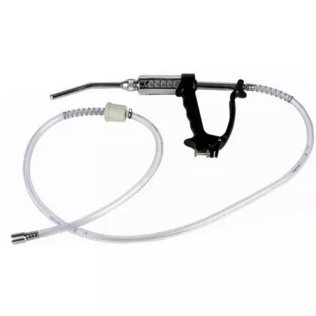
- Administration of Levamisole in individual doses of 7.5 mg / kg to pregnant sows, one week before farrowing, orally.
- Administration of feed medicated with Fenbendazole between 120 and 130 days old at a dose of 150 ppm.
With these indications the deworming program was strengthened, with special attention being paid to effective drug administration days before farrowing. The active ingredient in the antiparasitic was also changed to one that contains 0.2%. Ivermectin. The ages of treatment remained the same.
Following an analysis of the results we came up with 3 hypotheses.
1. Underdosing antiparasitic medication in the feed or to the sow days before farrowing.
2. Acquisition of resistance by the parasite.
3. Incorrect design scheme, very low number of samples.
Outcome of the case
After 7 months, the slaughterhouse reports the appearance of less scarring and prevalence of lesions and showed the trend in recent months to be a gradual decrease in the number of seizures. According to the veterinary inspector of the slaughterhouse, incidences are very low compared to when the case first appeared, although it is very important to mention that there are some weekly batches that arrive with an even larger number of affected pigs. The latter leads us to conclude that there are eggs in the beds even in the finishing buidings, some of which differ from one other.
Comments
Before commenting on the case report, we will summarize the life cycle of Ascaris suum. The nematode cycle begins in the egg and then enters orally as larvae migrating to the liver, lungs, and ultimately into the trachea. The animal swallows the larvae which reach the intestine to develop into the adult stage, a process that takes about 21 days, and once they are adults there is a 60 to 80 day delay in starting to lay eggs (Figure 6).
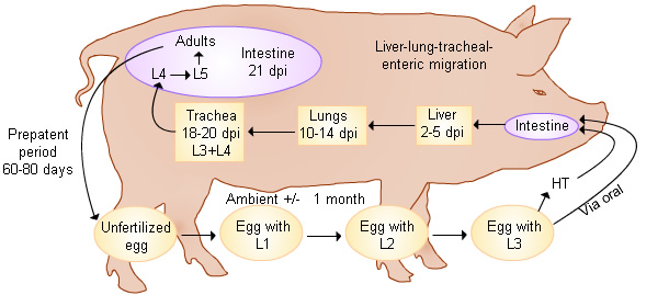
Figure 6. Development cycle of Ascaris suum.
The most likely hypothesis is that there has been anti-parasitic resistance in sows treated days before birth and in the feed of pigs. Also, the number of samples obtained were not representative in order to detect the presence of eggs.
The dynamics of parasite infestation should have been in different forms in the pig population. Ingestion of eggs during lactation is likely, since parasite control in sows is very difficult; the manual administration of antiparasitics is subject to a number of factors that may affect the effectiveness of product management, creating groups of farrowings where eggs are being secreted in high concentrations in the farrowing crates. However, if we take care that the female consume the product correctly and at the right dose, disease control is effective.
This case proves that the control of Ascaris suum on straw bedding must be considered very important because an imbalance in pig population health can occur at any time. Straw is highly favorable for the maintenance of eggs and their elimination is very difficult to accomplish. Additionally it is very important to make periodic changes of the antiparasite medication in order to prevent resistances.


