Description of the case report
In this case report we will describe an acute outbreak of gastric ulcers that affected 38 farms pertaining to a cooperative that produces compound feed in the area of Lleida (a province with a high swine density in the northeast of Spain) throughout a period of 12 months.

The first cases were diagnosed in October 2008, affecting, at the same time, various swine companies in Catalonia, Aragon and Valencia.
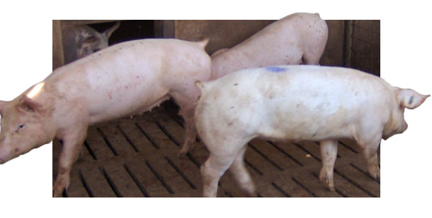
The only thing that all the cases had in common was the age of the affected pigs (30-40 Kg), the intensity of the outbreaks and the clinical symptomatology. Otherwise, the differences linked to the installations, genetics, handling, companies and compound feeds made it more difficult to connect the cases to a common cause. The severity and the level of losses ranged between a 5% and the 40% in the most serious cases.
Clinical symptomatology and necropsies
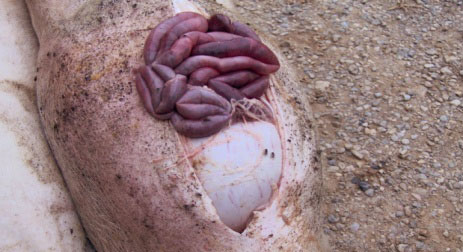
The only symptomatology seen was a mild paleness in the animals some 16 hours before the sudden appearance of the losses. The most severely affected animals were, in most cases, the biggest ones in the pen and they did not show other signs of disease. With the first losses, the presence of confusion and lethargy in a high number of animals in the affected buildings was common.
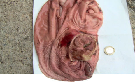
The necropsies carried out showed an acute gastric haemorrhage with a great stomach enlargement, mesenteric oedema, big-sized ulcers and, in an 80% of the cases, a severe hepatomegaly, an orange coloration and a friable consistency of the liver.
First measures taken
As the components of the compound feed, as well as its presentation were suspects of being the culprits, the first actions carried out were on the formulation:
- Increase of the particle size from 700 to 800 µm
- Decrease of the incorporation of wheat from a 45% to a 25%
- Incorporation of soya husks at a level of a 2% as a fiber source
- Incorporation of bicarbonate at a level of 10 Kg/Tm
- Increase of the levels of Vitamin E and Selenium (Se) at a level of 40 and 0.5 ppm, respectively
- Dietary compound feed in the most affected farms in the form of meal.
Laboratory results
After all these changes, the outbreaks persisted and their seriousness was the same. It was then decided to send macroscopically affected livers and apparently normal livers to the Unit of Anatomical Pathology of the Autonomous University of Barcelona (UAB).
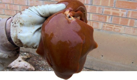

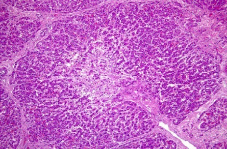
Histologically, a centrilobular necrosis of the hepatocytes and a massive destruction of the liver structure with mononuclear inflammation and megalocytes was found. These lesions were compatible with a chronic intoxication and/or a hepatosis dietetica.
Investigation of the possible causes
With the diagnosed liver lesion we started to consider different hypotheses as the primary cause of the appearance of the ulcers. All of them were related, initially, with possible toxic substances for the liver compatible with the lesion and with the toxic nature of the laboratory diagnosis.
First hypothesis: Suspicion of an aflatoxicosis
Among the many possible toxic substances that can harm the liver, micotoxins are the most probable ones in animal feeding.
We looked for aflatoxins, deoxynivalenol, zearalenone, ochratoxin, fumonisins and tricothecenes in the raw materials and the compound feed by means of HPLC in different specialized laboratories. The result was negative or positive, but at low levels.
Even so, in the case of mycotoxns it is very difficult to carry out a representative sampling in the compound feed factory, so these results were not conclusive. It was then decided to look for the same mycotoxins in the affected livers by means of HPLC MS/MS in the Technical Laboratory Rotterdam. The results were negative too.
The theory of the mycotoxicosis as the primary cause of the ulcers was losing soundness. It also had another weak point: the compound feed factories of the company are multispecies, and the fowl production was not affected at all, and they are more sensitive than pigs with respect to certain mycotoxins.
Second hypothesis: Suspicion of a toxic substance
Due to the absence of results and the seriousness of the outbreaks, we suspected of some other stranger and less common toxic substance for the liver.
The toxic substances for the liver include hundreds of them, because the liver is the most important detoxifying organ in the body but, due to the possibility of the appearance in the compound feed, we looked for the following elements in the following international centers:
- Heavy metals in the liver, kidney and subcutaneous fat by means of optic spectrometry and atomic absorption (Eurofins Ltd.London)
- Pyrrolizidine alcaloids by means of HPLC in the compound feed (Lab. Quantum Barcelona)
- Herbicides and insecticides by means of HPLC in the compound feed and the liver (TLR Lab. Rotterdam)
- Toxicological study by means of thin layer chromatography in order to detect some kind of toxic substance for the liver (Toxicology Department of the UAB)
The only finding highlighted by all the aforementioned analyses was a slight peroxidation of the compound feeds, but it was not possible to establish if it was real or maybe due to the storage of the samples. No sign of the rest of the investigated toxic substances was found.
Third hypothesis: Suspicion of a Vitamin E deficiency
Seeing that nothing conclusive that could cause a chronic intoxication was found, we decided to investigate the other possible diagnosed cause: the hepatosis dietetica. Clinically speaking there was no symptomatology, and there were also no mulberry heart disease nor white muscle disease findings in the necropsies, but nevertheless the possibility was borne in mind and this was investigates more thoroughly.
An analysis of Vitamin E and Se in the compound feed, the compound feed, the blood serum and the blood was carried out.
Table 1. Vitamin E and Selenium levels in the blood serum.
| Serum | December 2008 | |
| Samples | Vit E (µg/ml) | Se (µg/ml) |
| 1 | < 0.5 | 0.93 |
| 2 | < 0.5 | 0.78 |
| 3 | < 0.5 | 0.73 |
| 4 | < 0.5 | 0.61 |
| Ref. | 8-10 | 0.4-0.8 |
The results were much under the minimum recommended levels.
We decided to treat all the affected fattening stages with injectable Vitamin E and Se (Sodium selenite 1.5 mg; Tocopheryl acetate 50 mg). The results were very favorable in an 85% of the treated fattening stages. We also increased the Vitamin E levels in the supplements of the piglets, the fattening pigs and the sows.
Conclusions and solution of the case report
Vitamin E is the most powerful natural antioxidant so, therefore, it is a protector of the membranes, acting against oxidation reactions of the cell in vivo, among other functions. It is a treatment of choice against liver cirrhosis in humans, so it could be acting as a healing treatment in this case too.
On the other hand, hepatosis dietetica can be accompanied with gastric ulcers. Even so, the hepatosis could not be replicated at an experimental level by feeding pigs from 30 to 100 kg live weight without Vitamin E nor Se.
The Vitamin E reserves in the liver can be altered by toxic substances, mycotoxins in general or peroxides resulting from rancidity processes. Even the vitamin E levels in the compound feed are reduced with the presence of these toxic substances.
An important factor to be highlighted was the age at the onset of the problems (from 30 to 40 kg live weight in all of the cases), being this the moment at which the pig expresses its growth potential at its maximum level, so it subjects its vital organs to a great stress.
At the end of year 2008, the prices of Vitamin E increased considerably at a worldwide level. The general trend was to reduce the inclusion of vitamin E in the supplements, but also... could the quality have been affected?, was there any toxic substance or mycotoxin in the compound feed that reduced the vitamin E availability in the body of the animal?, or, could this toxic substance subject the liver to such an oxidative stress that this organ needed higher levels of antioxidant in order to work properly?
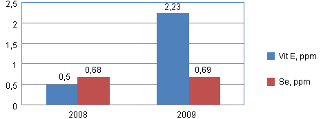
At the beginning of year 2009, the levels of vitamin E in the compound feed were increased. It was then analyzed again in the blood serum. Graph 1 shows the results of both analyses.
Graph 1. Comparison of the levels of vitamin E and Se in blood.
The authors of this article wish to consider as coauthors all the fellow veterinarians, workers and managers of the company that collaborated actively and helped during the case.


