Throughout the last decades, artificial insemination (AI) in swine has taken a radical turn with the addition of new methodologies such as post-cervical AI. In short, this type of AI consists of depositing the semen in the uterine body using a small diameter internal cannula, which runs along the inside of a traditional (cervical) insemination catheter. Despite the fact that this technique has shown an efficiency > 95% in multiparous sows (Watson and Behan, 2002; Bennemann et al. 2004), it has certain limitations, especially with regard to gilts. In fact, its successful application is lower in primiparous sows (~ 86%; Sbardella et al. 2014), and very reduced in nulliparous sows (~ 20%; Hernández-Caravaca et al. 2017.) All this suggests that the limitation of post-cervical AI in young females (especially in nulliparous) is due to morphological reasons and, therefore, associated with development, although no evidence has been reported to date. That is why we set out to make a comparative study of the reproductive system between nulliparous and multiparous sows, paying particular attention to the cervix, which is where the main problem of post-cervical AI lies. The problem is the difficulty to direct the internal cannula of the insemination device through the most cranial part of this organ. The study allowed us to check different significant aspects that confirm the existence of morphological differences. First, as one would expect, the length of the vaginal vestibule-vagina-cervix assembly is greater in multiparous than it is in nulliparous sows. In addition, the thickness of the cervical wall in the most cranial part of the cervix (near the body of the uterus) is higher in multiparous sows, but similar to that of nulliparous sows in the part closest to the vagina. In the same way, the composition of the tissue is different between both types of sows: the muscular tissue of nulliparous sows is proportionally bigger than that of multiparous sows, which suggests that the lower muscle tone in this area on sows, would facilitate the advance of the internal cannula during post-cervical AI. Finally, silicone moulds were obtained from the cervical canal, providing a very revealing visual information.
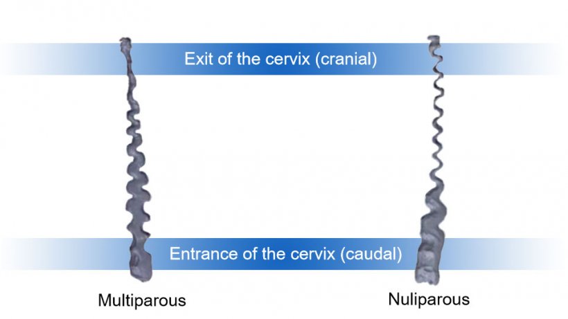

As Figure 1 shows, the differences found in the morphology of the cervical canal are significant, its diameter being noticeably bigger in multiparous sows, especially in the cranial end. Digital reconstructions of the cervical canal were made from these moulds (Figure 2).
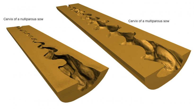
It is clear, therefore, that the morphological differences in the cervical wall and canal between these two types of sows, provides clarifying evidence that resolves the uncertainty about the current low applicability of the post-cervical AI in nulliparous sows. Hence, in this type of sows, the design of deep AI devices (those that go beyond the traditional cervical AI) must take into account the aforementioned morphological aspects. All these reasons lead us to test a new AI device adjusted and re-dimensioned to the particular anatomical characteristics of this type of sows. Due to the difficulty in crossing the entire cervix to reach the body of the uterus, as the anatomical study shows, a deep cervical seminal deposition is performed (Figure 4).

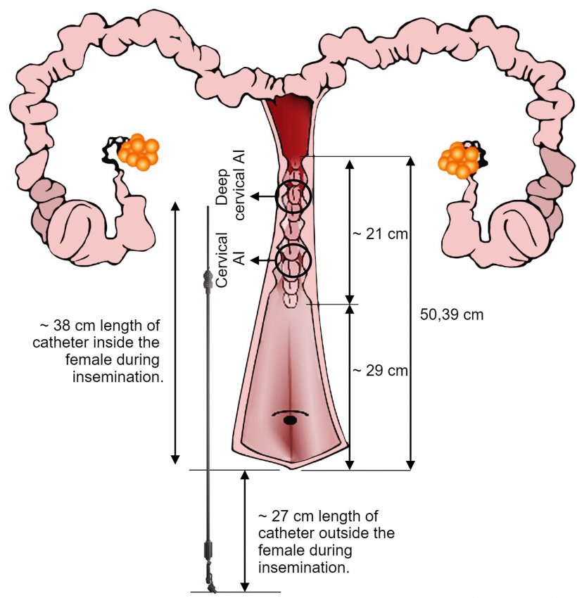
A field test was carried out with this new device, using more than 1,000 nulliparous sows (with 1 or 2 oestrus previously detected, 142.31 ± 8.27 kg live weight). Execution was successful in 89% of the inseminations. Subsequently, the reproductive data obtained after the AI (pregnancy and farrowing rates, number of piglets born) were compared with those of a cervical AI, where a lower number of sperm was used (deep cervical: 1,5×10⁹/45ml vs. cervical: 2.5 × 10⁹/85 ml). No significant differences were noted between both groups, which demonstrates the efficiency of the new technique.
The goal of these new studies is to broaden our knowledge of the physiology and anatomy of the swine female, in order to further improve AI and other aspects in this livestock species


