Mycoplasma hyopneumoniae (M hyo) causes chronic respiratory infection leading to significant economic losses due to lower growth performance and higher treatment costs. The overarching goal M hyo control programs is to minimize sow shedding. This reduces the number of positive piglets at weaning, which is a predictor for M hyo clinical disease in growing pigs.
A challenging aspect to achieve M hyo disease control is the acclimation process of incoming negative gilts into M hyo endemically infected farms.

How can we acclimate gilts at the farm?
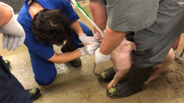
Traditionally, practitioners have attempted to expose negative gilts to M hyo through either direct contact with naturally infected animals or by intratracheal inoculations of farm-specific lung tissue homogenate, combined with a suitable vaccination and medication program. The issue with the former is that transmission of M hyo is known to be extremely low. In fact, a study carried out by Dr. Maria Pieter’s group, at the University of Minnesota, showed that the ratio of infected to naïve gilts should be 1:1, to guarantee infection of the naïve gilts in a 4-week period. This mean that achieving effective gilt acclimation through pig-to-pig contact would take a considerable amount of time and requires a large proportion of seeder gilts. On the other hand, the intratracheal inoculation, which is mainly carried out in research environments, requires skilled personnel, it is labor-intensive and it is a stressful procedure for the animals. Recently, veterinarians have explored the use of foggers to aerosolize M hyo into a confined air space to ensure gilt exposure through indirect contact (Yeske, 2019). Besides these methods and less-explored approach is intranasal inoculation.
Table 1. Summary of the advantages and disadvantages of each of the most common inoculation methods.
| Intratracheal | Intranasal | Aerosol | |
|---|---|---|---|
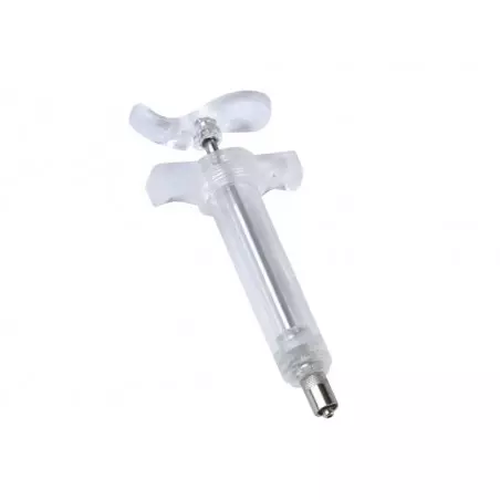
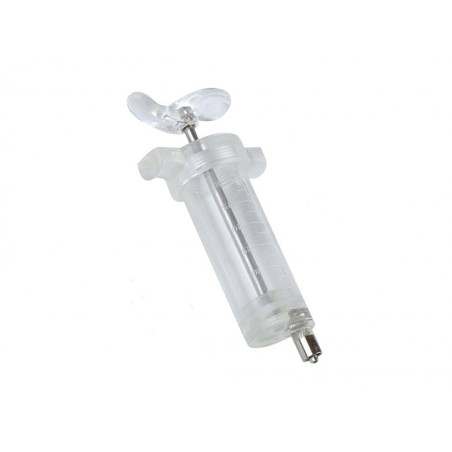
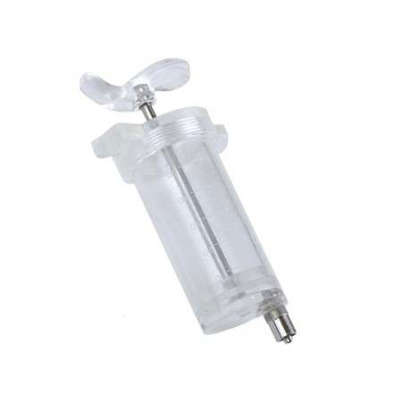
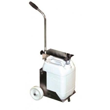
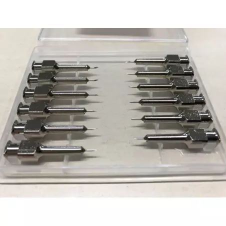
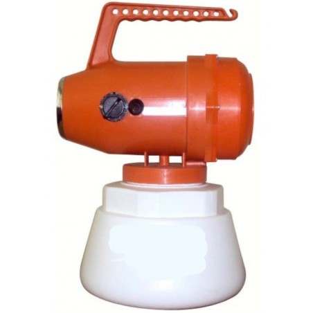
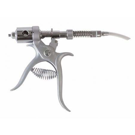
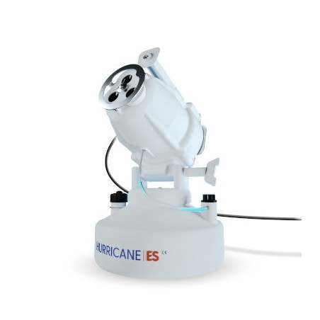
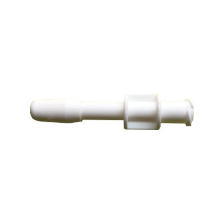
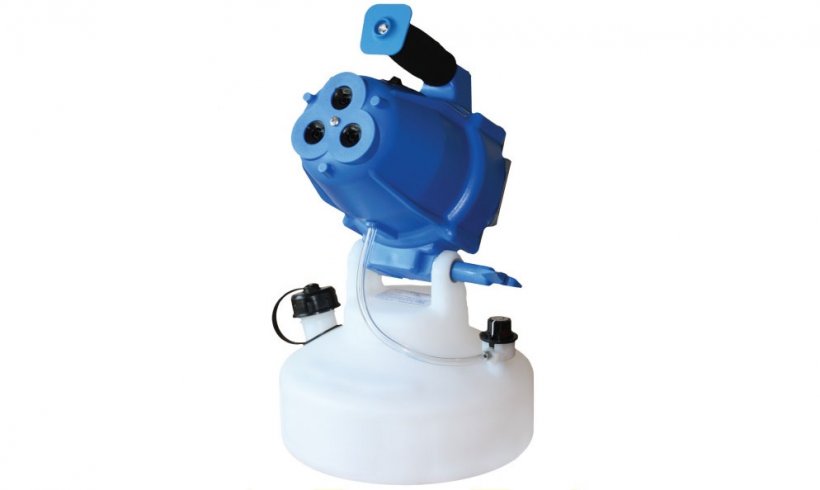
| Materials | Snare, mouth speculum, laryngoscope, catheter | Mucosal atomization device (Figure 1). | Hurricane fogger (Figure 3). |
| Application | Individually per animal | Individually per animal | Pooled per room or pen |
| Cost | Around $0.20 to $4 per device (disposable item) | Around $12 to $16 per device (can inoculate multiple animals with one device) | From $300 to $500 (non-disposable item) |
| Perform | Pros: Well-known and validated method. Generate consistent exposure of gilts |
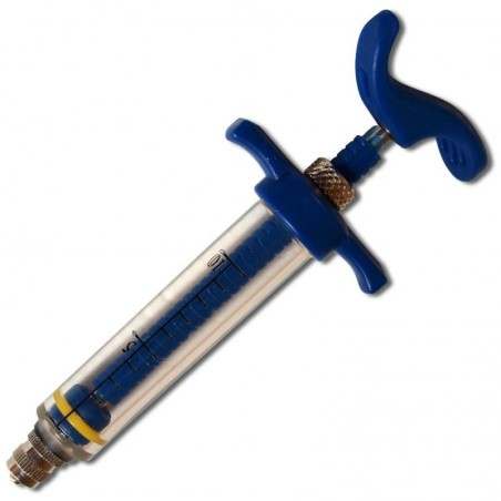
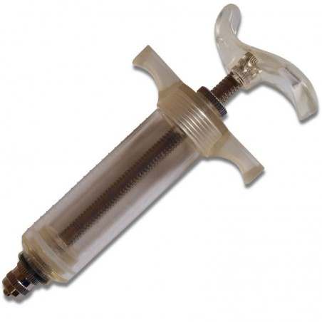
Pros: Less labor intense than intratracheal exposure. The device attaches to any luer-lock syringe, making it easy to implement on farm. |
Pros: Do not need skilled personnel. Same device can be reused. Requires the use of protective equipment for personnel No animal handling Can expose a large group og gilts in a short time |
| Cons: Need skilled and trained personnel. Labor and time intense Stress on pigs due to handling |
Cons: Need skilled and trained personnel. |
Cons: Require validation of procedures (i.e. dose, and time of aerosolization). Require appropriate farm conditions (i.e. facilities, ventilation, layout, size). |

How can we help farmers decide which is the best method?
Due to the different options, a group of researchers at Iowa State University and field practitioners collaborated in a recent study to address such challenge. The objective was to compare the efficacy of three inoculation methods (intratracheal, intranasal and aerosol) of live M hyo lung homogenate for practical, safe and reproducible gilt acclimation.
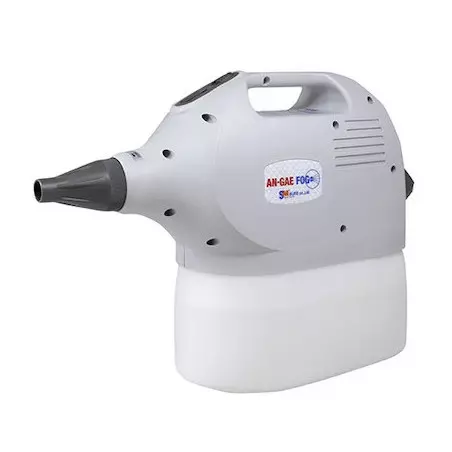
Six-week-old M hyo- and PRRSV-negative gilts (n = 78) were randomized to one of four M hyo exposure groups [aerosol (n = 24); intranasal (n = 24); intratracheal (n = 24); or no exposure (n = 6)] and followed through 49 days post exposure (DPE). The M hyo inoculum consisted of lung homogenate (105 CFU/mL M hyo 232). For the aerosol group, a cold mist fogger was utilized to aerosolize the lung homogenate. For the intranasal group, a malleable atomization device was used to administrate half of the inoculum volume into each nostril (5 ml) for two consecutive days. This device is extensively used in human medicine for pediatric nasal and tracheal drug administration in children. Weight was taken at -1, 28, and 49 DPE. Serum and tracheal samples for antibody and DNA testing were collected weekly through DPE 49. Pigs were humanely euthanized at 49 DPE. Lungs were evaluated for gross lesions and by histopathology (H&E and IHC). Weight gain, antibody responses (ELISA S/P), and shedding (qPCR) were statistically compared.
Results
All routes of exposure resulted in infection. Intratracheal demonstrated earlier detection of DNA (7 DPE) and seroconversion (39 DPE); intranasal and aerosol showed similar time to DNA and antibody detection, i.e., ~14 DPE and ~49 DPE. MHP qPCR Cts were similar in exposed groups over DPE (p>0.05), but lower ELISA S/Ps were observed in intranasal and aerosol exposures. Aerosol group had the least impact on daily gain (0.64 kg/day), performing similarly to the negative control group (0.73 kg/day). At 49 DPE, gross and/or histopathologic lesions were observed in the lungs of 17/22, 19/24 and 19/24 gilts from the intratracheal, intranasal and aerosol groups, respectively).
Take home
While intratracheal exposure resulted in the earliest detection of DNA and antibody, successful infection was achieved by all routes. Intranasal and aerosol routes might be better alternatives compared to the more labor-intensive and invasive intratracheal inoculation. Nonetheless, specific production circumstances may affect the choice of route. Practical and efficacious gilt exposure methods will improve M hyo control and elimination programs.


