Introduction
The case is based on a commercial 350 farrow to finish sow farm located in Northern Ireland.
Gilts are homebred in the unit. Boars are used for teasing. Gilts and sows are serviced with artificial insemination from a single source.

The farm purchases the creep and link feed (between 10 and 20 kg BW). However, it home mixes all the other feed rations.
History
The slaughterhouse contacted the farmer due to suspected animal welfare issues from the high number of healed broken ribs during the meat inspection of the carcasses.
The farmer contacted the veterinary surgeon of the pig unit for a second opinion, as a consequence of the slaughterhouse complain.
Investigation
Clinical Investigation
A visit to the slaughterhouse was carried out in September 2023 by the farm’s veterinary surgeon. Results from the slaughterhouse inspection assessment are summarised in Table 1.
Table 1. Results from slaughterhouse examination.
| Total animals slaughtered | 103 |
|---|---|
| Number of animals examined | 92 |
| Average enzootic pneumonia score | 1.3 |
| % Lungs with no enzootic pneumonia lesions | 90.2 |
| % Pleurisy | 14.1 |
| Average pleurisy score | 0.4 |
| % Necrotising pneumonia | 4.3 |
| % Lung abscesses | 4.3 |
| % Pericarditis | 5.4 |
| % Milk spot | 6.5 |
| % Mange | 9.8 |
| % Tail biting | 0 |
| % Broken ribs | 83.7 |
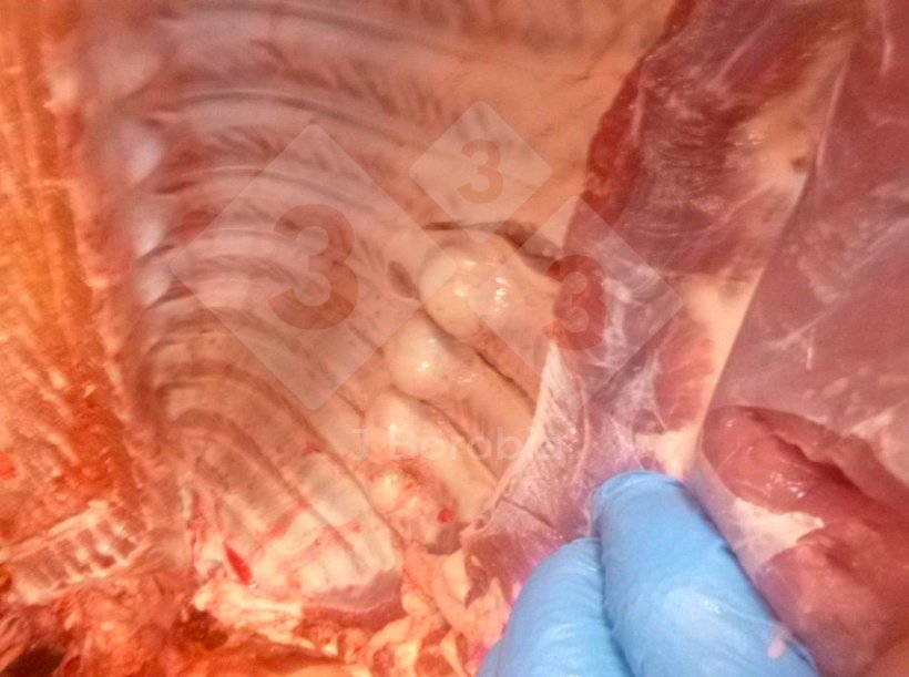
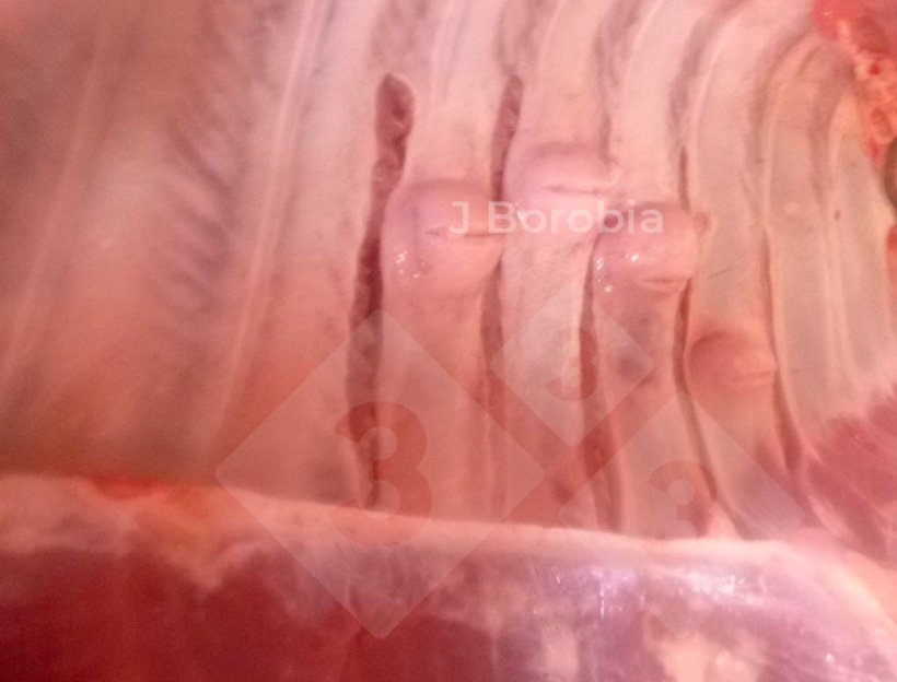
There were 77 carcasses with midshaft bony exostoses in the ribs (Figures 1 & 2). This number represented 83.7% of the carcasses inspected during the slaughterhouse visit.

Figure 1. Midshaft bony exostoses on the ribs of a slaughtered pig.

Figure 2. Midshaft bony exostoses on the ribs of a slaughtered pig.
Animals were clinically examined on the farm. No lameness or visible abnormalities were identified.
Stocking densities in the unit were within the normal parameters suggested by the Code of Practice Issued under the Welfare of Animals Act (Northern Ireland) 2011.
Mortality from weaning to finishing was 5.2%. The main cause of mortality was related to enteric disorders post-weaning.
A dead finishing pig was necropsied on the farm. Midshaft bony exostoses were detected in the ribs (Figure 3).

Figure 3. Midshaft bony exostoses on the ribs of dead pig on-farm.
Laboratory Investigations

Biochemistry was carried out on the blood of three 22-week-old pigs. The results are summarised in Table 2.
Table 2. Blood results from biochemistry. (Figures outside the normal range values are in bold).
| Calcium (mM) | Magnesium (mM) | Phosphorous (mM) |
Creatine Phosphokinase (U/l) |
Glutathione Peroxidase (U/g Hb) |
Vitamin E (uM) | |
|---|---|---|---|---|---|---|
| Pig 1 | 1.95 | 1.05 | 3.42 | 4956 | 288 | 9.1 |
| Pig 2 | 1.88 | 1.14 | 2.74 | 2828 | 283 | 9.8 |
| Pig 3 | 1.90 | 1.21 | 2.58 | 4579 | 337 | 4.8 |
| Lower Limit Upper Limit |
1.9 2.9 |
0.5 1.2 |
1.6 3.4 |
0 5000 |
> 2.3 |
Bone tissue (vertebrae and rib bones) was collected from the carcasses of four slaughtered animals with bony exostoses and analyzed for their mineral contents. The results are summarised in Table 3.
Table 3. Tissue results from biochemistry of the bone (vertebrae and ribs). (Figures outside the normal range values are in bold).
| Calcium (%) | Magnesium (%) | Phosphorus (%) | Ash (%) | |
|---|---|---|---|---|
| Pig 1 | 22.2 | 0.58 | 18.6 | 23.9 |
| Pig 2 | 28.5 | 0.58 | 19.1 | 18.6 |
| Pig 3 | 27.0 | 0.61 | 19.2 | 24.1 |
| Pig 4 | 32.5 | 0.83 | 18.6 | 27.0 |
| Normal values | 37 - 40 | >0.5 | 17 - 19 | >50 |
Nutritional Investigations
Based on the bony exostoses found on the ribs during post-mortem examination on-farm and in the slaughterhouse and the biochemistry results from Tables 2 & 3, diets were examined.
Due to financial problems, the farmer didn’t include the recommended mineral premix in the ration for six months.
Differential diagnosis
Based on all the investigations, infectious causes were ruled out.
Other possible causes of midshaft bony exostoses considered initially were:
- Overstocking: Stocking densities on the farm were within the normal requirements.
- Rough handling of pigs on the farm: Pigs were not handled roughly on the farm. There were far too many pigs with bone lesions and without associated bruising to indicate trauma.
- Rough transport: The lesions were chronic in nature. No recent bone fractures were present.
- Nutritional deficiencies.
A diagnosis of nutritional deficiency of calcium: phosphorus resulting in poor mineralisation of the bone leading to rickets and fibrous osteodystrophy in growing pigs was made based on the investigations.
Treatment program
The mineral mix was added to the feed mix with immediate effect.
Response to the treatment program
Factory reports indicated a result of a reduction of bony exostoses in slaughtered pigs six weeks after reformulating the diets. No more bony exostoses were detected in slaughtered pigs in a subsequent inspection in the slaughterhouse three months later.
Discussion
With modern dietary formulations, clinical deficiencies arising due to defective diets are unusual. Problems can occur due to faulty storage, the incorrect application of the feed, or interactions that reduce the availability of nutrients to the pig.
Calcium contributes to a wide variety of functions in the body including, but not limited to, blood clotting, muscle and nervous activity, hormone production, milk production, growth, and bone development. However, its major role together with phosphorous is in the formation of bone. Calcium forms approximately 38% of the bone structure with a further phosphorous 20% (Muirhead et al., 1997).
According to Banks (1981) and Burkitt et al. (1993) the bone is a specialized connective tissue composed of 65 – 70% of inorganic salts (mainly hydroxyapatite crystals – Ca10(PO4)6(OH)2 – and other elements in minor measure such as cations – Mg, Mn, Na,… – and anions – lactate, citrate, carbonate, Cl, F –) and 30 – 35% of organic matrix, osteoid (± 90% collagen type I and the rest are proteoglycans – chondroitin sulfate & hyaluronic acid – and non-collagen proteins – osteocalcin, osteonectin, sialoproteins, and other proteins –).
The finding of multiple carcasses with bony exostoses in the ribs led to the biochemical and nutritional investigations. It became apparent that a metabolic disorder related to the diet may have contributed to the problems experienced in this farm by causing decreased mineralization of the bone.
Rickets are a failure of mineralization of bone and growth plate cartilage (metaphyses of the long bones) in young growing pigs. It usually takes 6 to 8 weeks before symptoms become evident, by which time the disease has progressed to become irreversible (Muirhead et al., 1997). The most common causes are dietary insufficiencies of phosphorus or vitamin D. Calcium deficiencies can also cause rickets, and while this rarely occurs naturally, poorly balanced diets deficient in calcium have been said to cause the disease. As in most diets causing osteodystrophies, the abnormal calcium: phosphorus ratio is most likely the cause (Grünberg, 2022).
Pigs are sensitive to rickets development because of rapid growth rate and limited exposure to sunlight in modern confinement facilities (Dittmer, 2011). Pigs also have a tendency to develop fibrous osteodystrophy when faced with metabolic disorders, and this can occur concurrently with rickets as a secondary response (Thompson et al., 1989 & Thompson, 2007).
Reference ranges for analysis of bone ash have described 37 to 39.5% as the normal percentage value for calcium (of bone ash) and 19.6 to 20.6% as the normal percentage value for phosphorous (of bone ash). The ratio of Ca:P in the bones analyzed was affected. Moreover, the overall percentage of bone ash was below 50%. This decreased mineralization of the bone may be responsible for the bony exostoses observed in the ribs. Bone mineral assays in Done et al. (1988) cases showed lower levels than normal of calcium and phosphorous. Blood magnesium and calcium were within the reference limits.
The addition of a properly formulated mineral bag in the home mix diet had a positive effect in eliminating this problem.


