According to the current EU legislation on animal welfare and protection of pigs (2008/120/EC), tail docking should not be a routine practice. In order to encourage the transition to raising pigs with long tails, it is recommended to start raising subgroups of pigs with intact tails. This allows personnel to be trained, minimizing losses in the event of outbreaks of tail biting, which are unfortunately unpredictable in some cases. With this in mind, a conventional 1800-place finishing barn with all-in-all-out management brought in a finishing herd with a group of 300 piglets with intact tails. The average weight of the animals upon entry was approximately 33 kg. The floor is slatted and 2 feedings per day are provided. Upon entry, the animals showed no clinical pathological signs, neither enteric nor respiratory, and no locomotor or lameness problems were observed. However, among the 300 pigs with long tails, approximately 15% had scabbing and occasional signs of tail inflammation (predominantly redness and swelling, with slight tenderness to touch). Most lesions were resolving and no fresh blood was observed, although several animals had shortened tail length. The group of animals with intact tails was housed in separate pens from the animals with docked tails.
The pens were equipped with straw dispensers, used as environmental enrichment material available at all times. In addition, each pen was equipped with a metal chain attached to one of the perimeter walls.

"Deferred" losses
No signs of tail biting had been observed in pigs with intact tails immediately after entry and the healing process of previous lesions continued until completely resolved in the following weeks. However, about 5 weeks after entry, some animals began to show signs of lameness in the hindquarters, initially subtle and then gradually becoming more and more evident. Over the next 4 weeks, 22% of the animals that initially showed a tail lesion (9 animals), which then healed, had progressively lost the use of the hind limbs until they could no longer walk.
An animal with severe tail lesions resulting in paralysis of the hind limbs. On this same farm, the same clinical picture has been observed in animals in which the initial tail lesion was already healed and not visible.
Myelitis and ascending infection
The locomotor symptoms of animals that had previously presented tail lesions were accompanied by ascending infections to the sacral and lumbar vertebrae, often also with the presence of abscesses and myelitis, found during necropsy. The previous presence of lesions on the tail had constituted a route of entry and infection by secondary pathogens, whose activity had continued to ascend even after the superficial skin lesion had healed. The infection had resulted in the involvement of nerve structures and total or partial paralysis or paresis of the hind limbs.
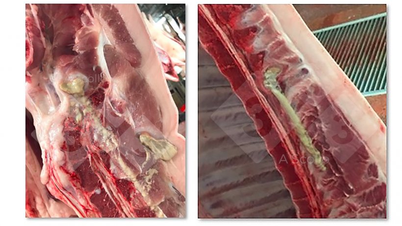
The problem with pigs on the farm that lose the ability to walk is certainly related to animal welfare, but also with non-negligible economic losses. In total, the loss of animals in the intact tail group was 7.4% compared to 4.9% in the rest of the tail-docked batch. In the intact tail group, losses were due to animals that could not be loaded on the truck due to the inability to walk on their own and were therefore euthanized on the farm, and to carcasses being seized at the slaughterhouse due to abscesses in the sacral area and viscera. In addition, the intact tail group saw an increase in expenses related to medication usage from €0.98 to €1.58.
Comparative summary between tail-docked animals and animals with intact tails.
| Group with intact tails | Group with docked tails | |
|---|---|---|
| Mortality | 7.4 % | 4.9 % |
| Myelitis (% of total mortality) | 22 % | 0 % |
| Cost of medications | 1.58 € | 0.98 € |
| Slaughterhouse losses | 4 % | 0 % |
Description of lesions
The clinical picture was frequently associated with the presence of secondary lesions in the spine, characterized by direct or compressive lesions on the distal motor neurons responsible for the mobility of the hind limbs. Authors (Hariharan et al, 1992) who have performed bacterial cultures of such lesions have often failed to isolate any pathogen, probably due to the chronicity of the lesion itself or the tendency to repeatedly treat animals with this condition with antibiotics. Macroscopically, the tail lesions were characterized by loss of the distal portion and possible scarring after scab detachment. Below the scar, there was often purulent material that was also in the intradural space up to the lumbosacral segment of the spine.
Increased risk of paralysis in case of tail biting
The proportion of animals with infections associated with tail-bite lesions (suppurative arthritis, suppurative spondylitis, and ascending bacterial meningitis/myelitis) increases with tail-biting events. This is also confirmed by a recent study (Piva et al, 2022) conducted by a Brazilian research group. In fact, these infections can even affect almost 70% of animals with bitten tails, while they represent only 0.04% of cases when the tail is not damaged. Simplifying, the risk of occurrence of these infections with clinical paresis or "delayed" hind limb paralysis after tail biting events is 57 times higher than in groups of animals without tail lesions.
Prevention is more important than treatment
Unfortunately, it is well known that it is difficult to intervene in the event of a tail-biting outbreak on pig farms. However, it is equally difficult to stop the risk of ascending infections after a severe tail injury. The risk may remain even if the injury is partially or completely healed, as the damage to deeper or visceral structures may be even greater than that exclusively affecting the skin. The "delayed" appearance of locomotor symptoms with respect to the original lesion is linked to the chronicity of an initially acute inflammatory and infectious condition, and to the spread of a pathogen from one tissue to another which can take so long that it makes it difficult to attribute it to the triggering event. The culmination of the process is, therefore, the clinical sign in a place different from the original one, sometimes associated with systemic lesions and, therefore, not always easy to interpret. One of the most complicated aspects of this process is also the fact that many times the animal continues to eat correctly and continues to grow, delaying the time at which the staff opts for euthanasia, prolonging the state of discomfort of an animal that can no longer walk and at the same time worsening the economic losses associated with an animal that has a poor prognosis continuing to consume feed. Prevention of tail biting events, by reducing environmental and management risk factors, is certainly the most useful intervention in these cases, with the understanding that animals with recent and evident lesions should be treated individually with antibiotic therapy to reduce the likelihood of infection.


