Why disease derived from exposure to aerosols containing M. hyopneumoniae is of importance?
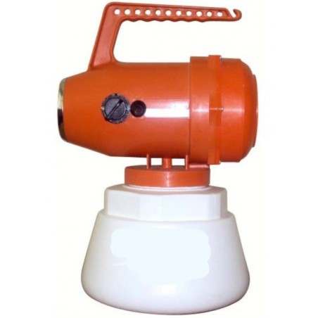
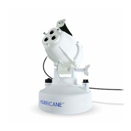
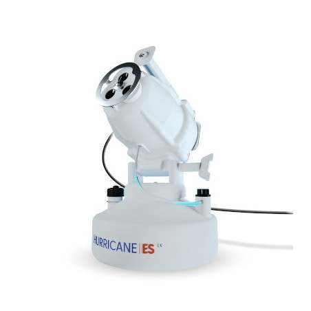
Controlled and artificial exposure of gilts to lung tissue homogenate containing Mycoplasma hyopneumoniae (M. hyopneumoniae) via aerosol has been proposed as a gilt acclimation strategy in the field, mainly in the United States. This strategy aims at reducing the presence of positive gilts at farrowing and therefore avoid infection of the progeny (Pieters and Fano, 2016). In addition, M. hyopneumoniae elimination programs are also commonly applied, which may include the aerosolization of medium containing lung tissue positive to the pathogen to achieve population exposure prior to start (McDowel et al., 2023). Hence, the infectivity and clinical course of disease derived from aerosols of M. hyopneumoniae is of current interest.
Understanding disease dynamics associated with M. hyopneumoniae exposure via aerosol
Since first described in 1965, M. hyopneumoniae experimental models of infection have been widely used to study different aspects of the disease as well as to assess vaccine and antibiotic efficacy. While intratracheal is the most widely used inoculation route for infection, aerosol is the least used inoculation method despite its similarity with natural infection (Garcia-Morante et al., 2017b).

Artificial exposure to M. hyopneumoniae via aerosol closely mimics the route of transmission thought to be the most important in naturally occurring infection.
With the aim of developing and characterizing an aerosol model for reproduction of mycoplasmal pneumonia in pigs, an experiment was carried out to determine the pathogenicity, colonization, mucosal immune response, and clinical course of disease of dose-controlled aerosols of M. hyopneumoniae.
Experimental design
Four groups of three M. hyopneumoniae-free gilts each were individually exposed to aerosols of diluted lung homogenate containing M. hyopneumoniae strain 232 in a chamber (Figure 1). Each group was exposed to different doses (Table 1).
- Nasal, laryngeal, and deep-tracheal secretions were collected from each gilt at 0, 7, 14, 21, and 28 days post-exposure (dpe).
- Blood samples were collected at 0 and 28 dpe to assess seroconversion.
- At necropsy:
- Lung lesions were assessed.
- Bronchial secretions and bronchoalveolar lavage fluid (BALF) were collected from each lung set.
To evaluate mucosal IgG and IgA production a modified ELISA was performed in BALF, deep-tracheal and nasal secretions.
To evaluate bacterial load a real-time PCR was done in nasal, laryngeal, deep-tracheal, and bronchial secretion samples.
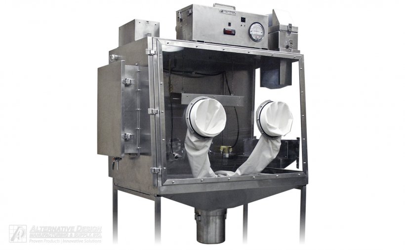
Table 1. Experimental groups and exposure conditions. Aerosol exposure was conducted once on two consecutive days (day 0 and 1) with M. hyopneumoniae strain 232.
| Experimental group | Titer | Total volume | Exposure time |
|---|---|---|---|
| LD/SE | 105 CCU/mL | 10 mL | 15-20 min/day |
| LD/LE | 105 CCU/mL | 20 mL | 30-35 min/day |
| HD/SE | 106 CCU/mL | 10 mL | 15-20 min/day |
| HD/LE | 106 CCU/mL | 20 mL | 30-35 min/day |
LD/SE= low dose/short exposure; LD/LE= low dose/long exposure; HD/SE= high dose/short exposure; HD/LE= high dose/long exposure; CCU= color changing units.
Results
M. hyopneumoniae naive status was confirmed in all gilts via the absence of antibodies and lack of detection of the pathogen prior to challenge. Thereafter, M. hyopneumoniae was detected by real-time PCR in pigs as early as 7 dpe in various sample types.
- All gilts in this study became infected and mean bacterial loads in different sample types did not differ substantially between experimental groups.
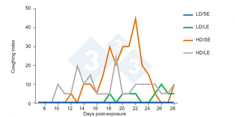
- The clinical presentation of respiratory disease varied according to the exposure dose of M. hyopneumoniae. Coughing was notable in the high-dose groups, whereas it was imperceptible or poorly detected in low-dose groups (Figure 2).
- All gilts from high-dose groups developed mycoplasmal pneumonia, whereas pneumonia occurrence and severity were lower in the low-dose groups.
- Consistent specific local humoral immune response (IgA and IgG) in deep-tracheal secretions was observed from 21 dpe onwards, regardless of the experimental group.
Reproduction of mycoplasmal pneumonia by aerosolization was successfully achieved
Since all gilts were exposed under the same conditions to the same M. hyopneumoniae strain, the present results suggest that the inoculum dose impacted the clinical infection outcome more than infection dynamics or mucosal humoral immune response.
Gilts became infected at approximately the same time, regardless the infectious dose
Conclusions and implications
Experimental models of disease are an indispensable tool to assess pathogenesis, dynamics of the immune response, and efficacy and safety for potential innovative therapies. In this study, the clinical and pathological manifestations, the infection and immune response establishment showed that the reproduction of mycoplasmal pneumonia by aerosolization could be successfully achieved, without inferiority in relation to other more classical inoculation routes, such as the intratracheal inoculation. These routes, in addition, require animal restraining and are less practical in the field.
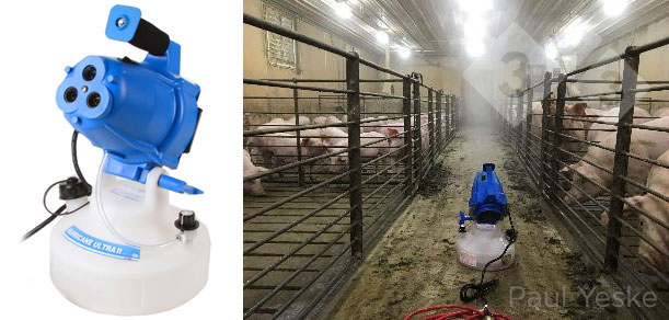
The ability to establish M. hyopneumoniae infections by aerosols may allow several types of experiments to be performed that are otherwise not possible or that are confounded when more artificial inoculation routes are used. In addition, the existing knowledge gaps on the pathogenesis of the naturally occurring condition, and the use of fogging systems for M. hyopneumoniae gilt acclimation or elimination programs, justify the development of a disease model using aerosols.


