Introduction
Gastro-intestinal disorders in pigs are extremely common and are often multifactorial, especially post-weaning. In the author’s experience, post-weaning diarrhoea will occur at some time or another on more than 50% of pig farms. It is therefore clinically important to try to reach as accurate a diagnosis as possible of specific causes in order to apply appropriate control measures, which might include nutritional and management changes, vaccination, antimicrobial therapy and other supportive measures. This is sometimes difficult because of the common occurrence of mixed infections, the compounding effects of environment and nutrition and the difficulties experienced in isolating and identifying all the possible pathogenic agents involved.


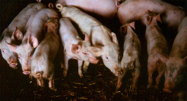
Weaned pigs with diarrhoea, showing varying degrees of weight loss and dehydration.
Differential diagnosis of enteric disease in pigs
The presence of enteric disease in the herd is usually obvious. In determining the causes, it is important to assess the full clinical picture, taking into account all factors that might be relevant. This would include details of recent pig movements onto the premises and herd biosecurity protocols, since several enteric pathogens such as salmonella, rotaviruses and porcine epidemic diarrhoea virus can be introduced by carrier pigs. The clinical features of an outbreak will usually alert the clinician to the most likely cause or causes, which can be confirmed if required in the diagnostic laboratory. Samples to be submitted in an investigation might include fresh feces, intestinal or rectal swabs and ligated loops of bowel for bacteriology, virology and parasitology; freshly-fixed sections of intestine for histopathology; and blood for serology. Other samples might include feed and drinking water for bacterial culture and for assays of a variety of substances including mycotoxins, antimicrobials, heavy metals, etc.; samples of feces from rats, mice and birds can help identify vectors; swabs taken from various points in buildings and vehicles can help identify how infection is being spread and where it might be harboured. Proper sampling protocols and many samples are required if the environmental pathogen burden is to be assessed.
Examination post mortem of a representative sample of pigs that have died on the farm can also provide valuable diagnostic information. The gross pathological lesions of many of the enteric disorders of pigs are regionalised and are visually quite striking. The main differential diagnoses are shown in Table 1.
Table 1. Guidelines for differential diagnosis of main causes of post-weaning diarrhoea in pigs
| Nature of diarrhoea | Gross pathology and site of lesions | Clinical picture | Possible causes | Further tests |
| Watery, alkaline. Grey-brown | - Ileum. Thin walled, distended, prominent. - Peyer’s Patches. - Enlarged mesenteric lymph nodes, may be haemorrhagic. |
- Spreading infection within contact group. - Increasing mortality. - Weight loss. - Dehydration. |
- E. coli - Rotavirus |
- Culture. - Haemolysis. - Demonstration of F4 and F18 fimbrial antigen. |
| - Oedema of stomach, mesentery and peri-orbital region. | - Acute. High mortality in affected pigs. Vocal changes. | - E. coli oedema disease. | ||
| Watery, flecks of necrotic material. Yellow-brown | - Ileum congested. - Haemorrhagic lymph nodes. - Polyserositis. - Catarrhal enteritis. - Diptheritic membrane. - Prominent Peyers Patches |
- Spreading infection within contact group. - Pyrexia. |
- E. coli - Salmonella |
- Culture and serotyping. |
| Creamy, grey-brown, possibly some blood | - Ileum congested. - Spiral colon congested and firm. - Haemorrhagic mesenteric lymph nodes. |
- Limited numbers within groups. - “Sudden” deaths without diarrhoea. |
- C. perfringens Type A - Brachyspira spp. |
- Culture. - Enterotoxin profile. |
| Profuse, watery, green-yellow | Ileum thin walled and distended. | - Rapidly spreading through groups. - Rising mortality. |
- TGEv - PEDv | - Virus isolation. |
| Grey-dark grey-brown. Maybe projectile | - Non-specific. Some congestion of small and large intestine. - Foul smelling. - Maybe gastric ulceration. |
- Widespread, sudden onset (nutritional) otherwise gradual spread. - Low mortality. - Weight loss. - Maybe catarrhal or haemorrhagic inflammation of terminal section of ileum. |
- Nutritional - Antibiotic perturbation of gut flora. - L. intracellularis - B. pilosicoli |
- Rule out infectious causes. - Culture and PCR - Serology for Lawsonia. |
| Grey-dark grey-brown. Maybe blood |
- Large intestine necrotic, haemorrhagic. - Colon contents may contain purple-tinged blood.- Necrotic smell. |
- Slow spread within groups and then between groups. - Chronic weight loss. - Rising mortality. |
- Brachyspira spp., especially B. hyodysenteriae. | - Culture and PCR. |
Infectious causes of diarrhoea in fattening pigs

The author’s experience is that the most common infectious causes of diarrhoea soon after weaning are Salmonella enterica (S. typhimurium) and E. coli, particularly types possessing fimbrial antigens F4 (K88) and F18. Lawsonia intracellularis, Clostridium perfringens and Brachyspira pilosicoli can be implicated and, occasionally, Brachyspira hyodysenteriae. Rotavirus is not uncommon and from time to time persistent diarrhoea after weaning as a result of gut damage due to coccidiosis pre-weaning is encountered. We are fortunate in the UK that porcine epidemic diarrhoea is not a problem at the time of writing.
Non-infectious causes of diarrhoea in fattening pigs
Any sudden change in feed can precipitate dietary upset but the effects are usually of short duration. Pigs in the early phases of growth, up to around 25kg liveweight, tend to be less tolerant of feed ingredients of low digestibility and thus in this age group, feed specification is more critical. Profound physiological changes occur as a result of abrupt weaning, notably a change for high pH to low pH in the stomach; a change in the profile of digestive enzymes, particularly to deal with starch; and a shift in the recovery of water from the foregut to the hindgut. At the same time, stunting of the intestinal villi reduces absorptive capacity in the ileum. These changes render the gut more susceptible to pathogens in the short term through changes in substrates which may lead to abnormal fermentation patterns.
There is a strong association between the presence of gastric ulceration and finely ground diets. Feeding a 550 µm diet seems to maintain or slightly aggravate abnormalities in the pars oesophageal epithelium, but a 750 µm diet allows healing. The use of a structure mill as opposed to a hammer mill gives greater uniformity of particle size as well as larger particles and experience in the field suggests that a change away from hammer milling significantly reduces the incidence of gastric ulceration and diarrhoea. Gastric ulceration also occurs post weaning when piglets have erratic feed intake or they do not actually eat at all in the first 24-48 hours after weaning. Furthermore, this can give rise to perturbation of the normal gut flora.
Excessive use of antibiotics for a variety of conditions can also cause serious upset of the gut flora and, in some cases almost sterilise the gut, allowing the proliferation of abnormal flora such as yeasts. This leads to a persistent diarrhoea, often exacerbated by the administration of further course of antibiotics in a futile attempt to control a situation precipitated by antibiotic treatment in the first place. The prolonged use of sulphonamides for the control of Strep. suis or Haemophilus parasuis can select resistant strains of E. coli and Salmonella, which can then lead to enteric symptoms that are more difficult to control.
Poor storage and hygiene conditions can lead to oxidation of fats, the development of mycotoxins and accidental contamination with many substances, all of which may precipitate or aggravate diarrhoea.
Antinutrient factors in raw materials that may precipitate episodic diarrhoea include saponins, phytates, lectins and trypsin inhibitors but the mechanisms remain poorly understood.


