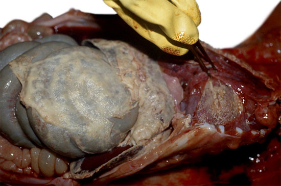
Fig. 1. Pig with Glässer’s disease. Noticeable presence of fibrin in the peritoneal cavity (fibrinous peritonitis) and pericardiac cavity (fibrinous pericarditis).
In 1995 and 1997, Vahle et al inoculated by via intranasal animals with H. parasuis isolated from a pericardiac lesion, and they studied the development with time of clinical signs, lesions and organs from which the bacterium was isolated. In these experiments, after the intranasal inoculation, the bacterium could be recovered from the lung before blood or any other systemic organ and before the typical lesions of Glässer’s disease developed.
These results indicate that H. parasuis, after entering the animal through the respiratory system, has to reach the lung and survive the pulmonary defences before it can invade the blood stream, and from there it reaches different organs where it can multiply and produce the inflammation which is typical of fibrinous polyserositis and arthritis, characteristics of Glässer’s disease.
Furthermore, H. parasuis is, together with other bacteria such as Streptococcus suis, one of the early colonisers of piglets. As a coloniser, H. parasuis can be isolated from the upper respiratory tract of healthy piglets in which it does not produce any pathology. Therefore it is normal to isolate distinct strains of H. parasuis on a farm or even in a single animal. However, under certain circumstances this bacterium can produce serious infections. Apart from Glässer’s disease, H. parasuis can produce catarrhal purulent bronchopneumonia and septicaemia without polyserositis.

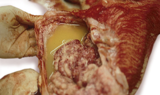
Fig. 2. Abdominal cavity of a pig with Glässer’s disease. In some cases the fibrinous exudation is accompanied by a large quantity of a yellowish liquid. This liquid is a very adequate sample for the isolation of the bacterium.
The infections from H. parasuis are spread across all countries that produce swine and they cause important economic losses mainly due to post-weaning deaths and slow growth in the chronically affected piglets. Historically, Glässer’s disease has presented itself in a sporadic manner in piglets of 1-4 months age that have been submitted to stress. In traditional production systems, piglets receive the bacterium from their mothers during suckling, at the same time as they receive maternal immunity which offers them protection. In this way the piglets are colonised while they are protected by the maternal immunity, until they develop their own antibodies against the prevalent strains on the farm. However, the production systems have been modified and the introduction of early weaning and specific pathogen free (SPF) animals or of high sanitary status farms have provoked an increase in the prevalence and severity of many infections including those produced by H. parasuis.


