The porcine respiratory disease complex is still one of the most challenging issues in the pig industry worldwide, and enzootic pneumonia (EP)-like lesions, characterized by cranioventral pulmonary consolidation and pleuritis are the most frequent findings in pig lungs at slaughter. For this reason the abattoir is a useful endpoint to collect data on herd health status and a valuable tool for monitoring the effect of disease control measures.
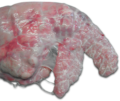
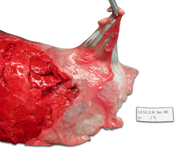
In particular, chronic pleural lesions (CP) are commonly detected at the abattoir, since the resolution of pleuritis can take 3 months or more and very often the process is not completed prior to slaughter (Andreasen et al., 2000). The most important cause of pleuritis in pigs is Actinobacillus pleuropneumoniae, but several other bacterial pathogens are also involved, particularly Haemophilus parasuis, Pasteurella multocida and Streptococcus suis (Meyns et al., 2011). The localization of pleuritis can be cranioventral or dorsocaudal (fig. 1 and 2) and the border between the two areas is marked by the dorsal limits of the interlobar fissures. Cranioventral pleural lesions are strongly associated with complicated EP-like lesions, whilst dorsocaudal lesions are considered suggestive of lungs that have recovered from A. pleuropneumoniae (Christensen and Enoe, 1999). The Slaughterhouse Pleuritis Evaluation System (SPES) made by Dottori et al. in 2007, shows five different scores (Table 1) depending on the extension and location of the pleural adherences and provides two major outputs: (1) The SPES average value, i.e. the sum of each lung score/number of scored lungs, and (2) the A. pleuropneumoniae index (APPI), i.e. the frequency of pleuritis lesions with a SPES score ≥ 2 in a batch multiplied by the mean pleuritis lesion score of the animals with SPES ≥ 2.

 |
 |
| Figure 1: Right lung of a pig. Chronic ventro-cranial pleuritis involving the cardiac lobe and the cranial part of the diaphragmatic lobe. | Figure 2: Left lung of a pig. Chronic dorso-caudal pleuritis involving the cranial part of the diaphragmatic lobe. Characteristic stripping of the pleura. |
Table 1: The SPES score grid for chronic pleuritis (CP).
| Score | Lesion characterization |
| 0 | Absence of CP lesions |
| 1 | Ventro-cranial lesion: pleural adherence between lobes or at ventral border of lobes |
| 2 | Dorso-caudal monolateral focal lesion |
| 3 | Bilateral type 2 lesion or extended monolateral lesion (at least 1/3 of a diaphragmatic lobe) |
| 4 | Severely extended bilateral lesion (at least 1/3 of both diaphragmatic lobes) |
From April to June 2008 a total of 48 batches (100 pigs per batch on average) of heavy pigs (160 kg slaughter weight, aged 9–10 months) were scored at the slaughterhouse for chronic pleuritis (CP) using the SPES grid.
For each herd 20 blood samples were collected from 80 kg pigs and 20 additional blood samples were collected at slaughter for each batch. A. pleuropneumoniae seroprevalence was evaluated.
A total of 4889 lungs were examined. Chronic pleuritis (SPES score 1) was recorded in 2,322 (47.5%) lungs. Dorsocaudal pleuritis (SPES score ≥ 2), suggestive of recovered pleuropneumonia, was found in 1,227 lungs (25.1%). The mean SPES value of all the lungs was 0.83 (95% CI 0.78–0.86), ranging from 0.04 to 1.87. The mean APPI of all the studied batches was 0.61 (95% CI 0.51–0.71), ranging from 0 to 1.84.
These results showed that pleural lesions were frequently detected in slaughter pigs in Italy (47.5% of scored lungs) as was dorsocaudal pleuritis suggestive of previous A. pleuropneumoniae infection (25.1%). These high prevalences are in accordance with previous findings published by other authors elsewhere in Europe (Cleveland-Nielsen et al., 2002; Pagot et al., 2007; Marois et al., 2008; Meyns et al., 2011), although it is almost twice as high as that recorded in Spain (Fraile et al., 2010).
The SPES average value, the APPI and the percentage of lungs with SPES score ≥ 2 were associated with A. pleuropneumoniae herd seroprevalence, confirming the importance of this pathogen as a causative agent of CP. Two herds that were seronegative for A. pleuropneumoniae and that did not show dorsocaudal pleural lesions have confirmed the reliability of the SPES method. No statistical correlation was demonstrated between A. pleuropneumoniae seroprevalence and the percentage of lungs with SPES score = 1 (ventrocranial pleuritis), confirming the hypothesis that only dorsocaudal CP lesions are suggestive of recovered pleuropneumonia. In order to have data for ranking a batch with respect to the general population, from February 2008 to January 2011, lungs from 14,195 pigs belonging to 139 batches were evaluated using the SPES score grid. Forty-two per cent of the lungs showed chronic pleuritis (SPES score ≥ 1). Dorso-caudal pleuritis (SPES score ≥ 2) was found in 24% of the lungs. Lesions with score 2 were assessed in 14.3% and lesions scoring 3 and 4 were present in 8.3% and 1.6% of pigs, respectively. The mean SPES value of the overall lungs was 0.77. The mean APPI of all batches was 0.60. The APPI values were divided in four categories: best quarter < 0.28; intermediate best quarter from 0.28 to 0.53; intermediate worst quarter from 0.53 to 0.81 and worst quarter > 0.81 (fig. 3). The distribution in classes of the APPI values obtained in this study is used as a tool for ranking a batch with respect to the general population.

Figure 3: Distribution the of APPI values in four categories: APPI < 0.28 (best quarter); APPI from 0.28 to 0.53 (intermediate best quarter); APPI from 0.53 to 0.81 (intermediate worst quarter); APPI > 0.81 (worst quarter).


