Diarrhoea in growing-finishing pigs is a common problem in commercial farms and significantly limits the efficiency and profitability of swine production globally. Intestinal disorders in pigs older than 4 weeks of age can be produced by several infectious agents such as rotaviruses, coronaviruses, Escherichia coli, Brachyspira spp., Salmonella spp., Yersinia spp., Lawsonia intracellularis, Oesophagostomum dentatum and Trichuris suis, among others. In fact, most cases featuring postweaning diarrhoea and colitis are due to concurrent aetiologies, together with its interaction with feed, water and other environmental factors.
Porcine circovirus type 2 (PCV2), the essential causal agent of PCV2-systemic disease (PCV2-SD) and PCV2-subclinical infection (PCV2-SI), is considered a systemic pathogen. However, recently, it has been suggested as a pathogen able to induce diarrhoea in pigs, and PCV2-enteric disease (PCV2-ED) as a separate entity within porcine circovirus diseases (PCVDs) has been proposed. Its case definition includes: a) pigs suffering from diarrhoea; b) granulomatous enteritis and lymphocyte depletion observed microscopically in Peyer’s patches, but not in other lymphoid tissues, and c) moderate to high amounts of PCV2 detected in intestinal mucosa or Peyer’s patches without detection in other systemic tissues.

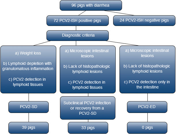
In order to clarify the true occurrence of PCV2-ED, a study was designed to investigate the prevalence of PCV2 infection in growing-to-finishing pigs with diarrhoea in Spain and assess the potential overlap between PCV-ED and PCV2-SD. The study was conducted in 96 pigs suffering from diarrhoea and submitted for complete necropsy and laboratory analyses to the Servei de Diagnòstic de PatologiaVeterinària at the Veterinary School of the Universitat Autònoma de Barcelona (Spain). Submissions (Fig. 1) corresponded to the period between 1998 and 2011, and included only non-PCV2 vaccinated pigs. The most prevalent enteric lesions were catarrhal (77.1%), fibrinous (11.5%) and granulomatous (4.2%) enteritis/colitis; other lesions such as haemorrhages or ulceration were also seen (4.2%). Seventy-two pigs (75%) were positive for PCV2 by in situ hybridization (ISH). Among positive pigs for PCV2 ISH, 39 animals were confirmed as suffering from PCV2-SD and 33 had no significant lymphoid lesions but low amount of viral nucleic acid in several lymphoid tissues. In consequence, these latter animals showed the existence of a systemic PCV2 infection and, therefore, did not qualify for PCVD-ED. In fact, the pathological interpretation of low amount of PCV2 several systemic lymph nodes with not significant histological lesions is potentially triple: a) PCV2-SI, b) initial phase of a subsequent PCV2-SD, or c) recovery phase of a PCV2-SD.

Fig. 1. Flow chart describing the selection and diagnostic criteria for PCV2 infected pigs included in this study.
PCV2, porcine circovirus type 2; PCV2-SD, PCV2-systemic disease; PCV2-ED, PCV2-enteric disease.
In conclusion, results obtained from this study (Fig. 1) suggested that PCV2-ED is probably a negligible condition and PCV2 mainly contributes to enteric clinical disorders in relation to PCV2-SD occurrence.
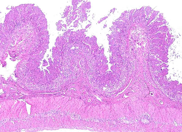
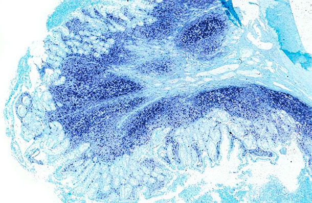
It is important to highlight that enteric problems were frequently found in PCV2-SD affected farms. However, the exact mechanism by which these affected animals showed diarrhoea was not clear. On one hand, the systemic immunosuppression linked to the PCV2-SD would be a major reason for triggering the effect of existing enteric pathogens in the farm, which may not be expressed unless such endangering of the immune system. Alternatively, the pathological outcome of PCV2-SD in a proportion of animals includes a variable degree of granulomatous enteritis (Pictures 1 and 2), which would cause diarrhoea due to the altered permeability of the intestinal barrier. In such scenario, the advent of PCV2 vaccines caused a radical decrease of pigs and farms displaying PCV2-SD clinical signs and lesions and, therefore, the concomitance of the viral infection with other enteric pathogens significantly decreased.

Picture 1. Ileum. Marked lymphocyte depletion and granulomatous inflammation of the Peyer’s patches at the ileum
of a pig affected by PCV2-systemic disease. Hematoxylin and eosin stain.

Picture 2. Ileum. The same ileum of the PCV2-systemic disease affected pig displays a massive amount of PCV2 genome
at the Peyer’s patches and the intestinal mucosa. In situ hybridization to detect PCV2; fast green counterstain.
Due to vaccination, the most common form of PCV2 outcome is the PCV2-SI nowadays, and systemic and/or digestive clinical signs and lesions caused by the virus are minimal. In consequence, when diarrhoea problems are present at an age in which PCV2-SI also occurs, it is very likely that concurrent infections with enteric pathogens take place and the latter ones are the main contributors to the clinical problem. Therefore, it is very important to use available diagnostic tools in order to ascertain the specific weight of each pathogen in the particular enteric disease. Pathological examinations (gross and histopathological) can be of great help trying to assess such scenarios.


