Enzootic Pneumonia (1/3): M.hyo, clinical signs and diagnosis
Christina Gale, BSc, Swine Marketing Manager, Eduardo Velazquez, MRCVS, Swine Veterinary Service Manager and Emma Pattison, BSc, Swine Field and Vaccination Services Manager, Ceva Animal Health, UK
The pathogen
Mycoplasma hyopneumoniae is 200-500nm in size and requires complex media and aerobic and microaerophilic conditions to be cultured. Infection with M.hyopneumoniae occurs via inhalation of infected aerosols or via direct contact with infected animals. The pathogen establishes itself in the respiratory tract, attaching to the ciliated cells of the tracheal, bronchial and bronchiolar epithelium. Once in the respiratory tract, it can persist for weeks to months and causes pathological changes in the lungs. Lesions are thought to form in the lobes of the lung due to lack of normal clearance of secretory products from the respiratory system, due to infection of the ciliated epithelium (Taylor, 1995). Early lesions are observed as small dark red areas in the anterior lobes, which enlarge over time and after a few weeks lose their red colour and become more pink (Figure 1). Lesions resolve after 12 to 14 weeks with formation of interlobular fissures (Maes et al., 2008). This is important to note, as infections early in production may be resolved once the animal reaches the slaughterhouse, so any scarring (Figure 2) should be noted as losses may still have occurred during the growing and finishing stages.
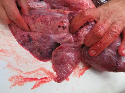
Figure 1 - Pneumonic lung lesions caused by infection with M.hyopneumoniae
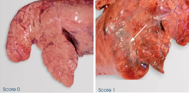
Figure 2 - Scarring of lobes (score 1) due to recovery of lung lesions caused by infection with M.hyopneumoniae
Histologically, inflammation occurs and neutrophils can accumulate in the airways and bronchioles. Lymphocytes and macrophages will also be present 5 days after infection, followed by presence of large mononuclear cells, polymorphs, lymphocytes and plasma cells in the alveoli from day 7 (Taylor, 1995). Immunoglobulin IgA forms in tracheal mucosa so may be detectable from day 30 on diagnostic testing of secretions, and IgG may also be present at a slightly later stage.
Clinical signs
Enzootic pneumonia can clinically be acute or chronic in its form which have different clinical presentations of disease. Pigs of all ages can be affected in the acute form and most likely will experience pyrexia, anorexia and respiratory distress, usually accompanied with a distinct cough (Taylor, 1995). Whereas, in the chronic form, few clinical signs may be observed, particularly in young growing pigs, where diarrhoea and a dry cough may be the only visible signs if present. Later in production, this cough may become a barking cough in the finishing house and what may be most apparent is the variation in size of pen mates, indicating performance has been compromised in some animals.
Diagnosis
Diagnosis can be carried out by M.hyopneumoniae specific seroconversion or laryngeal swabs tested by PCR (Pieters et al., 2017). Examination of the lungs at slaughter can also be useful to report presence of pneumonic lesions, characterised by consolidated areas especially in the cranial lobes (Maes et al., 2001). Seasonal patterns are often observed with M.hyopneumoniae, with lung lesions prevalence and severity increasing following the winter months. This was demonstrated by Ostanello et al. (2007) where, as scored using the Madec and Kobisch method (1982), mean lung lesion score was significantly increased in batches of animals raised in the winter months (October-March) compared to the summer months (April-September), with scores of 2.33 and 1.81 respectively. Decreased growth rate and feed conversion ratio (FCR) may also be observed, typically with no or low mortality (Sibila et al., 2009)

Click here to discover CEVA products!
Contact:
Contact us using the following form.


